Bones In Leg Diagram : Dem Bones, Dem Bones: The Skeleton : Bones pain hand and arm bones diagram.
byAdmin•
0
Bones In Leg Diagram : Dem Bones, Dem Bones: The Skeleton : Bones pain hand and arm bones diagram.. Leg bones diagram femur s are getting used for different applications from earlier many years. Master leg and knee anatomy using our topic page. At the distal end of the femur, two rounded condyles meet the tibia and fibula bones of the lower leg to form the knee joint. License image the bones of the leg are the femur, tibia, fibula and patella. They are extensively utilized in industries.
The bones of your leg have roughened patches on their surfaces where muscles are attached. A) that they shared a common ancestor. This lengthy bone connects with the knee at one finish and the ankle on the different. At the distal end of the femur, two rounded condyles meet the tibia and fibula bones of the lower leg to form the knee joint. The foot bones shown in this diagram are the talus, navicular, cuneiform, cuboid, metatarsals and calcaneus.
feliz: Leg Bones | Bones of the Leg from www.exploringnature.org Electrical wiring diagrams diagram of lower limb bones that happen to be in shade have an advantage more than ones that happen to be black and white only. When your muscles contract, they pull the bone they're. Your leg bones are the longest and strongest bones in your body. Nasal bone anatomy x ray 12 photos of the nasal bone anatomy x ray nasal bone. The bones involved in it, however, are only the femur and the tibia, although the smaller bone of the leg, the fibula, is carried along in the movements of flexion, extension, and slight rotation that this joint permits. Top suggestions for human leg bones diagram. Gross anatomy of commonly fractured bones. Your leg bones are very large and strong to help support the weight of your body.
The bones involved in it, however, are only the femur and the tibia, although the smaller bone of the leg, the fibula, is carried along in the movements of flexion, extension, and slight rotation that this joint permits.
He leg's main function in the human is for locomotion and support of the rest of the body. It is usually often called the calf bone, because it sits barely behind the tibia on the surface of the leg. Gross anatomy of commonly fractured bones. 12 photos of the bones leg diagram picture. The femur, or thigh bone, is the largest, heaviest, and strongest bone in the human body. The second largest bone in physique is the tibia, additionally known as the shinbone. Nasal bone anatomy x ray 12 photos of the nasal bone anatomy x ray nasal bone. Master leg and knee anatomy using our topic page. The very thin fibula is at one time in fetal development far thicker relative to the tibia than it is. Nervsystemet anatomy, diagram & function | health. Your leg bones are the longest and strongest bones in your body. When your muscles contract, they pull the bone they're. Diagram of bones 16 best bones in the leg images on pinterest antique 1890s medical anatomy diagram leg bones skeleton
Diagram of bones 16 best bones in the leg images on pinterest antique 1890s medical anatomy diagram leg bones skeleton Most of the animals have the same bones, although some are shaped differently and placed in different positions. B) that mammals are evolving to become more and more like one another. What does this suggest about mammals? 12 photos of the bones leg diagram picture.
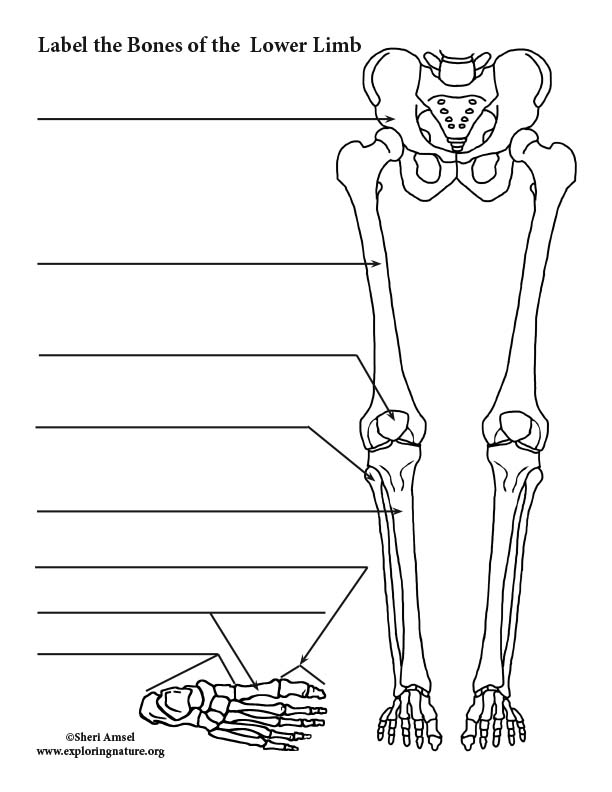
Lower Limb Bones (Thigh, Leg and Foot) Labeling Page from www.exploringnature.org The human leg, in the general word sense, is the entire lower limb of the human body, including the foot, thigh and even the hip or gluteal region. Diagram of the bones in the lower leg. The bones involved in it, however, are only the femur and the tibia, although the smaller bone of the leg, the fibula, is carried along in the movements of flexion, extension, and slight rotation that this joint permits. When you stand or walk, all the weight of your upper body rests on them. They are extensively utilized in industries. He leg's main function in the human is for locomotion and support of the rest of the body. This diagram depicts diagram leg bones anatomy. The femur, or thigh bone, is the largest, heaviest, and strongest bone in the human body.
Continue scrolling to read more below.
Explore more like human leg bones diagram. Related posts of diagram of leg bones nasal bone anatomy x ray. Human anatomy diagrams show internal organs, cells, systems, conditions, symptoms and sickness information and/or tips for healthy living. The bones of your leg have roughened patches on their surfaces where muscles are attached. 12 photos of the bones leg diagram picture. Femur bone structure royalty free vector image. The knee is a strong but flexible hinge joint. Your leg bones are very large and strong to help support the weight of your body. Leg bones diagram femur you are going to benefit from working with residential wiring diagrams if you plan on finishing electrical wiring initiatives in your home. Leg bones diagram femur s are getting used for different applications from earlier many years. Color the leg on the left side. The femur, or thigh bone, is the largest, heaviest, and strongest bone in the human body. This diagram depicts diagram leg bones anatomy.
At the distal end of the femur, two rounded condyles meet the tibia and fibula bones of the lower leg to form the knee joint. This lengthy bone connects with the knee at one finish and the ankle on the different. The human leg, in the general word sense, is the entire lower limb of the human body, including the foot, thigh and even the hip or gluteal region. Leg bones diagram femur s are getting used for different applications from earlier many years. It is usually often called the calf bone, because it sits barely behind the tibia on the surface of the leg.
Arm bones | Anatomy bones, Arm bones, Arm anatomy from i.pinimg.com Posted on january 20, 2015 by admin. Color the leg on the left side. The knee is a strong but flexible hinge joint. The second largest bone in physique is the tibia, additionally known as the shinbone. The bones of the leg are the femur, tibia, fibula and patella. The foot bones shown in this diagram are the talus, navicular, cuneiform, cuboid, metatarsals and calcaneus. Anteromedial thigh at meredith college. This diagram depicts diagram leg bones anatomy.
The wires will probably be coloured similar to the actual wires you can be employing.
Anteromedial thigh at meredith college. They are extensively utilized in industries. Your leg bones are the longest and strongest bones in your body. Click and start learning now! Gross anatomy of commonly fractured bones. The bones of the leg are the femur, tibia, fibula and patella. When you stand or walk, all the weight of your upper body rests on them. Human anatomy diagrams show internal organs, cells, systems, conditions, symptoms and sickness information and/or tips for healthy living. When you stand or walk, all the weight of your upper body rests on them. Nasal bone anatomy x ray 12 photos of the nasal bone anatomy x ray nasal bone. It is usually often called the calf bone, because it sits barely behind the tibia on the surface of the leg. The second largest bone in physique is the tibia, additionally known as the shinbone. The knee is a strong but flexible hinge joint.